Samuel Kim is a senior from Cupertino, California, studying neuroscience and psychology. While playing football in high school, he began his research at the Schmahl Research Institution in San Jose, California, to pursue his interest in traumatic brain injury (TBI) under Dr. Anuran Chatterjee. There, he developed a novel TBI model of tunable extracorporeal shock wave therapy in C. elegans with a manufacturing cost of $1. He went on to continue his work on TBI in the lab of Dr. Terrance Kummer in the department of neurology at the Washington University School of Medicine in St. Louis. For the past three years, he has worked on the development of a closed-head impact model of engineered rotational acceleration (modCHIMERA) and the subsequent super-resolution imaging and analysis technique, AirySynapse.
In the developed world, TBI is the number one cause of death and disability among young people (<45 years old). However, there is still no available treatment that we can offer beyond supportive aid. Therefore, understanding the pathophysiology that underlies TBI would benefit the development of potential therapies. In the Kummer lab, we are focused on creating the tools needed to help localize the foci of neural circuit dysfunction. Although the historical attention has been on axonal injury, with synapses below the resolution of classical techniques, new technology enables us to overcome this barrier.
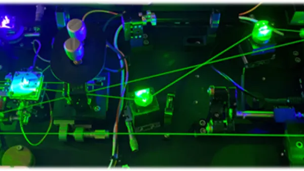
Synapses, the junctions between neurons that facilitate communication through chemical messengers, are a focus of injury in many neurological diseases. Through my Bio 500 and BioSURF experiences at WashU, I have worked extensively on an image analysis process we call AirySynapse, which facilitates the study of synapse loss after TBI. By leveraging advancements in confocal microscopy that exceed the diffraction limit of conventional microscopy, we are theoretically able to resolve 97% of all distinct synapses with the logistical ease and scanning capability of confocal microscopy.
By colocalizing pre- and post-synaptic markers, we can use colocalized puncta that are within biologically synaptic distances of each other as a proxy for putative synaptic density in a given volume. Utilizing this quantification method, we may easily identify synapse loss due to diseases such as TBI, Alzheimer’s, and dementia. Thanks to the scanning capability of confocal microscopy, we can compile mesoscale raster images of large brain regions to define these changes within their anatomical context, and then further characterize these synapses with respect to other elements of interest such as microglia and complement proteins through the use of additional channels. Moreover, this technique can be broadly applied to study any molecular subsets that feature biologically significant spatial relationships. We hope to continue to use AirySynapse as a tool to explain why synapses are degraded following TBI and what we can do to prevent their loss.
Though I originally arrived at WashU set on attending medical school, my experience in the Kummer lab through Bio 500 and the interactions with my professors and valued mentors have led me to reconsider. I am grateful to have realized that research is a stimulating process of continual growth that I want to pursue as a career, and I am hopeful for the findings that our generation of neuroscientists will discover about the complex organ that makes us human.