Inside a corner lab in the basement of Busch, thousands of worms lay in wait.
The windowless room stays at a cool 18 degrees Celsius. Small plastic containers (think the ones you might have put some craft supplies in as a kid) line the front wall, each a contained saltwater habitat where juvenile worms hide in tubes of their own creation.
It feels almost Marvel movie-esque—a multitude of worlds stacked on top of one another, where only a chosen few can see the full picture.
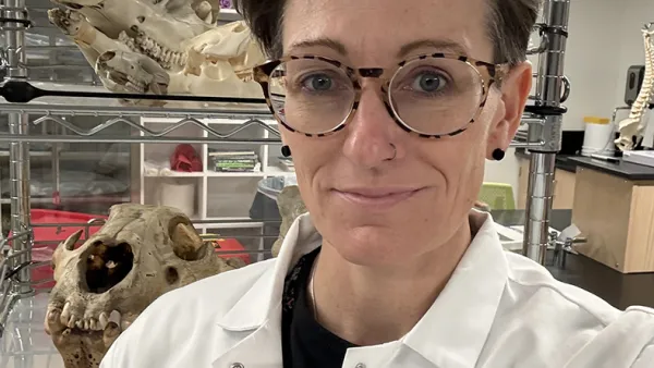
At the center of these universes is Duygu Özpolat, Assistant Professor of Biology. Her work revolves around the segmented worms—also called annelids—that crowd this room, looking at how they develop and regenerate. Think regrowth. Think stem cells. These are the research topics that everyone talks about, but these tiny creatures are often overlooked.

A day in the life
The worms—Platynereis dumerilii—may look small and simplistic to the naked eye, but under the microscope they are fully fledged beings—a mouth, eyes (two sets), legs, head, jaws. You can see blood pumping up and down their bodies, a flash of red in an otherwise neutral picture. Younger worms, the ones with just three or four segments, are, as Özpolat says, almost cute. They are nearly transparent under the microscope, useful for imaging cellular processes and structures under the skin. They look like you could give them a hug and they’d bounce right back. “Anyone who comes to the lab and doesn’t know these animals ends up falling in love with them,” Özpolat says.
Today, in this room of worlds, is mating day. The juveniles are separated by sex, but when the two groups finally meet, it’s as if there’s a sudden storm. Eggs and sperm cloudy the water. Females begin to wither and die (the males aren’t too far behind). And then, there it is—the beginning. “There are labs that study this particular species not for their regeneration, but for their quirky life cycle,” Özpolat says. “Their reproductive cycle is highly regulated by light. These animals synchronize their maturation—sexual maturation—based on the moonlight.” This moonlight is reproduced in the lab. An artificial romance. Still, it pays off: Thousands of eggs settle in the bottom of the container, so small that it’s hard to think anything of them.
Özpolat focuses the microscope on the egg-filled cup with ease, then offhandedly mentions that these types of eggs are everywhere in the ocean. “You’ve probably swallowed a few at some point,” she says.
I peer into the eyepiece of the microscope. Despite being nothing more than loose dots mere minutes ago, these tiny spherical beginnings are now encased by a jelly. They move together when jiggled. I ask what percentage of these eggs will hatch, expecting 50%, maybe 65%, but Özpolat responds with, “Most of them.” It surprises me, despite being surrounded by the evidence that these specks will one day be something more. And that one day will come very soon—today, in fact. Eggs hatch within twelve hours (compare this to the 2-3 weeks it takes for a tree frog egg to hatch). Then they will grow, and wait, for Özpolat to reach into their world once more.

A closer look at regeneration
These worms were a large part of the newest research from the Özpolat Lab published in Developmental Biology. This September issue also boasts a cover photo taken by postdoc Ryan Null—the worm is cream and spotted, almost otherworldly, against a sunset-colored background.
This study takes a closer look at how P. dumerilii regenerates body parts. “They regenerate all kinds of different tissues—nervous system cells, gut cells, simple kidneys. We’re particularly interested in their reproductive cell generation,” says Özpolat. Her team specifically focused on how maturity and age affect these worms’ ability to regrow reproductive cells when the bottom half of their body is amputated—a costly process in terms of energy.
The hypothesis here was obvious: researchers thought that the younger juveniles would be faster at regeneration than the older juveniles. “It’s hard not to think about ourselves,” Özpolat admits. She’s right that we often associate youth with resilience and faster healing. “We ended up having this bias towards thinking that older worms were going to struggle more or be slower at regenerating.” Surprisingly, the opposite was true for P. dumerilii. The older worms regrew their amputated parts and matured faster than their younger counterparts.
There is, as always, a catch: Older juveniles may be faster at regeneration, but only if they can survive the initial cut. And even when they do survive, the researchers noticed that they weren’t fully regenerated or that they were different than normal. The cover photo from Null depicts one such worm.
This research not only helps us better understand regeneration through different life stages, but also how energy is used by these worms as they grow and mature. Next, the Özpolat Lab wants to investigate the mechanistic details of hormonal regulation and how different hormones driven by the reproductive state of young or old worms translates into faster growth and regeneration. “Hormonal regulation is important to consider when studying regeneration, because it’s not just about where you cut, it’s about the entire animal and how it’s responding to that injury due to its reproductive state,” Özpolat explains.
Beyond this future research, Özpolat’s goal is clear: “I just want people to keep an open mind about worms. I feel like I’m on a side quest of getting more people to appreciate them.”


