Ivan Radin, a postdoc in the Haswell Lab, talks about his winning image.

Ivan Radin was born and raised in what is today Serbia. His exposure to science started at a young age through family members in various scientific fields. His mom and both maternal grandparents are/were university professors in the areas of microbiology and plant pathology. His dad runs a lab for seed quality control, and one of his paternal grandparents was a math and physics teacher, the other a pharmacist.
“Science and the love of nature were always part of my surroundings. My family never pressured me to go in one direction or another career-wise, but I caught the bug of scientific curiosity and fascination with nature early on, nevertheless. It might sound corny, but the thrill of discovering something new is truly exciting. I love how variable research is. There is always something new to learn or try,” Radin said.
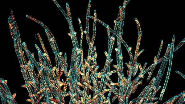
Radin, a postdoctoral fellow in the laboratory of Elizabeth Haswell, appreciates the welcoming atmosphere and diversity of people and research topics in the Biology Department at WashU. The Haswell Lab primarily studies plant mechanobiology, or how green organisms sense and respond to physical forces. Radin recently won the Olympus Image of the Year award for the region of Americas. His winning image features Physcomitrium patens, a type of moss used as a model organism in Haswell Lab’s research.

The cells on the bottom or closer to the center of the plant, are called chloronemal cells. They are smaller and have large chloroplasts that fill most of their interior. These cells are the main site of photosynthesis. The bigger cells that are spreading away from the plant are called caulonemal cells, and have smaller and fewer chloroplasts. These cells grow fast and explore new areas to expand the plant.
“It’s fascinating how without the context, the moss under the microscope almost looks like an alien. The image challenges our preconceptions of what the plant should look like. During my regular work, I do a lot of imaging and make hundreds of pictures trying to see structures or quantify something in order to answer a scientific question. However, every so often, a particularly beautiful scene or view comes across my eyepieces or screen, so I stop the experiment and take a few minutes to optimize the imaging conditions so I can take a nice picture of that scene. When inspiration strikes me, I follow it. Microscopy is a window into another world where we get to see beautiful and fascinating things that we’re normally unable to see,” Radin explained.
Radin also enjoys the challenge of making figures for scientific publications, because they need to be scientifically sound, clear, structured, and accessible (e.g., color blind friendly), yet also visually appealing and striking. He states that it can be quite a challenge to reconcile these sometimes-opposing requirements but that is also what makes it fun!
Read more about the Olympus Image of the Year competition and check out other winning photos here: https://www.olympus-lifescience.com/en/landing/ioty-2021/.
