Dianne Duncan talks about research in the Duncan Lab and the Biology Imaging Facility

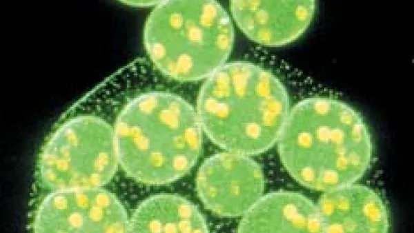
Dianne Duncan is originally from Pennsylvania. She received her undergrad degree in Biology at Penn State in 1981. She came to Washington University for graduate school in the then relatively new DBBS program. She finished her master’s degree in Developmental and Molecular Biology. The DBBS program was attractive and unique because it allows movement from one department to another. This flexibility provides students with a variety of options and experiences. Dianne’s thesis work was on the multi-cell alga Volvox in the lab of David Kirk.
After receiving her master's degree, she worked as a research technician in the lab of Ursula Goodenough from 1984-1988, until she joined Ian Duncan’s lab. Partners in life and work, they co-wrote grants and conducted research on genes that control pattern formation in Drosophila at many stages of development. This type of research has contributed a great deal to our knowledge of the genetic control of development in humans. The late 1980’s and early 1990’s were exciting times in this field of biology with so many discoveries regarding gene conservation between fruit flies and humans.
“It’s astonishing that the same gene in flies and humans is so interchangeable. What has become clear is that certain ‘master regulatory genes’ are conserved throughout evolution and that these genes work in the abstract; telling an organism what structure to develop. The organism then interprets this information in a way that is appropriate for its species. For a certain class of these genes, not only is the sequence conserved, but the order in which the genes in the complex are arranged on the chromosome has also been conserved throughout some 500 million years of evolution. The discovery and analysis of patterning genes led to Nobel prizes for three fly biologists,” Dianne said.
Current research: glowing grasshoppers and the referee gene
The Duncan Lab has been studying the E93 gene since 2010. Flies have four life stages. During the pupal stage, a number of structures are patterned such as wing veins, abdominal pigmentation and bristle structures. All normal pupal patterning events are disrupted in strong mutants of the E93 gene. What they have found is that the E93 gene product appears to act as a cofactor for many gene products to promote pupal (verses larval) patterning events.
“Imagine a soccer team. At half-time, the referee hands the players a new rule book, changing the game to field hockey. The players are the same, but the rules change the game. Like the referee, the E93 gene doesn’t appear at all until the pupal stage and then it’s everywhere acting as a cofactor to pattern everything in pupa. It’s a neat concept. The organism doesn’t need to have nearly as many genes when they can be re-purposed. This was a paradigm shift in how we look at how genes work,” Dianne explained.
The lab is studying how many genes E93 reacts with globally. They have isolated close to 40 genes (the data suggest there are hundreds more) that are direct responders to genes in flies.
The lab also provides transgenic glowing grasshoppers for a study by Barani Raman, at WUSM, who studies neural behavior in insects: https://sites.wustl.edu/neuronex/.
Biology Imaging Facility
Alongside her work in the Duncan Lab, Dianne also manages the Biology Imaging Facility, a core resource on the Danforth campus, used primarily by research labs in the Biology Department, but also by other departments on Danforth, including Anthropology, Chemistry, Physics, Engineering and some departments on the medical campus as well.
When she started managing the facility in 2010, all of the microscopes were running on different software. Over time, grants from the NSF and funds from the Biology Department led to gradually getting them all to match. Now that they all run on the same software, the processes are seamless and the same techs are able to maintain and service them, streamlining the whole system.
The facility is comprised of five rooms containing seven microscopes in the basement of McDonnell Hall (rooms 15, 16, 18, 19, 20). The newest addition from 2021 is the fancy Leica THUNDER Live Cell fluorescent compound microscope, which can image brightfield as well as blue, green, red and far-red fluorescent dyes at high resolution, and has a time-lapse function. This instrument can quickly tile over a whole slide, which works well for brain slice imaging and multi-well plates. Built-in de-convolution helps adjust for fuzzy light from the fluorescent samples that typically interfere with viewing the images.
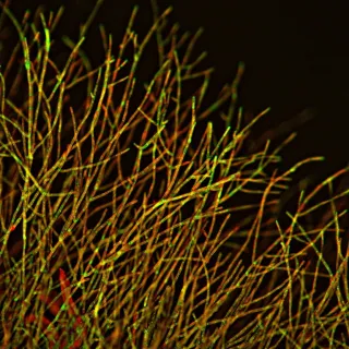
The Leica THUNDER Model Organism Macroscope was purchase by the department in 2019 and comprises a fluorescent motorized stereomicroscope for imaging color or multi-channel fluorescent images and movies of larger samples such as small plants, animals and insects.
The Haswell Lab and the Biology Department collaborated to purchase the Zeiss Evo10 Environmental Scanning Electron Microscope in 2016. This instrument provides stunning monochromatic images of sample surface features in magnifications ranging from 80X to 100,000X and more.
The Facility also houses three confocal microscopes. The oldest (2009) is the Nikon A1 Si Single Photon confocal microscope, which was the department’s flagship microscope for many years. To provide better speed and resolution, the Leica Sp8 Lightning Single Photon microscope was acquired in 2018. This instrument allows you to scan fluorescently labeled samples while mechanically removing out of focus light to generate sharp images at high resolution in three dimensions, and has become the most popular scope in the facility.
In 2018, faculty members Ram Dixit and Erik Herzog, along with Dianne, applied for a National Science Foundation Major Research instrumentation grant that resulted in the purchase of the Leica Sp8 DIVE Multiphoton confocal microscope. This instrument greatly increases the depth of imaging of fluorescently labelled samples; allowing for deep imaging of live and fixed animal brains as well as providing a suite of new technologies to probe living, fixed, labelled, and unlabelled samples at high resolution with very low phototoxity.
The facility also houses a small brightfield stereomicroscope with color camera, as well as a dedicated computer workstation for sophisticated post acquisition analysis of images obtained on any of the microscopes.
All microscopes and peripheral equipment within the facility are available for use by any researcher in the university community after training by Dianne. To learn more about the Biology Imaging Facility, the equipment, and how to make arrangements for use, visit https://sites.wustl.edu/imagingfacility/.

