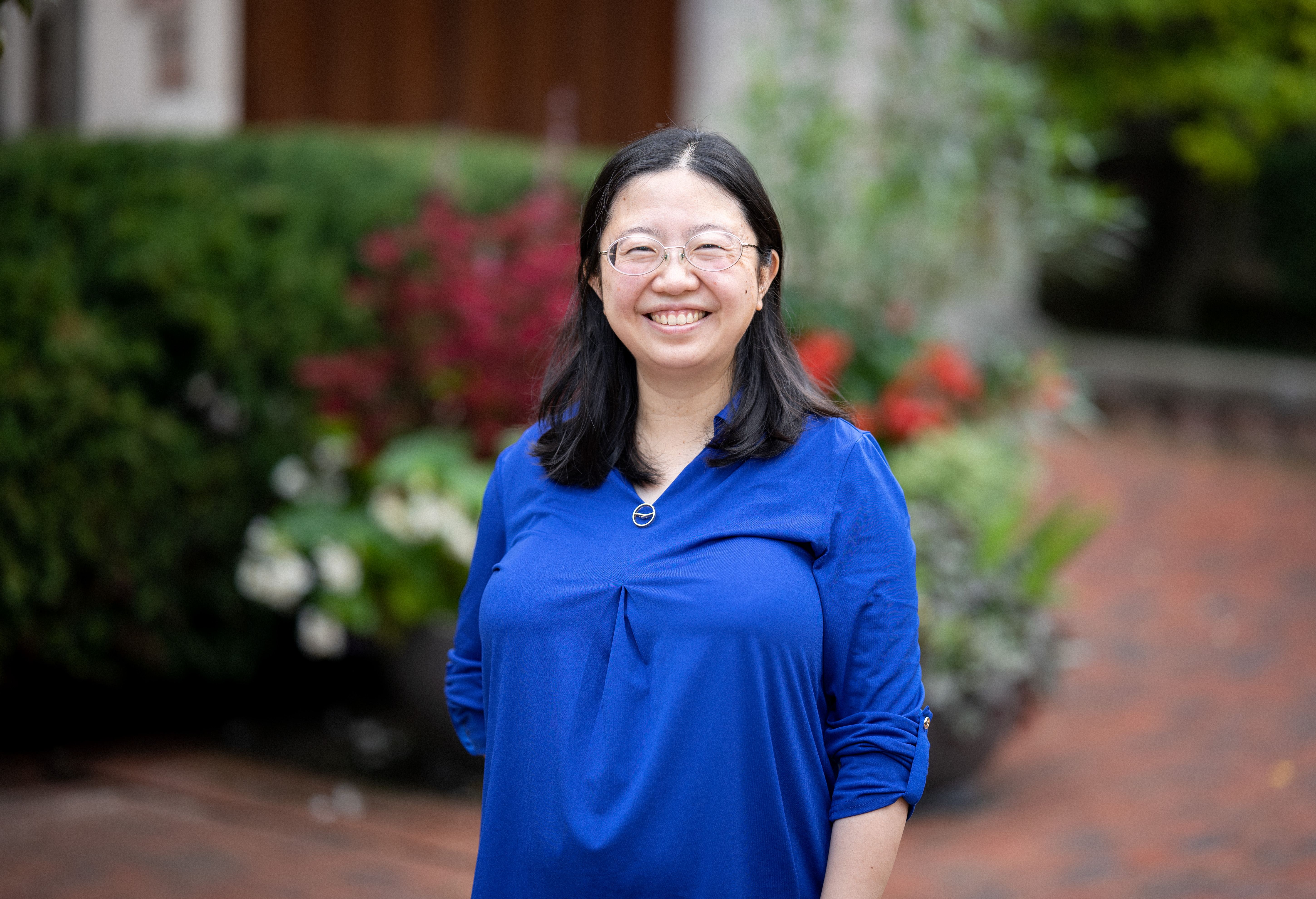
Jennifer Wang joined the Biology Department as Assistant Professor in July 2022.
Born in China, she came to the United States when she was 7 years old, and spent most of her childhood on Long Island, NY. She received her bachelor’s degree in Biology at MIT, after which, she decided she needed more research experience. She spent two years working as a research assistant in a lab at Harvard Medical School. Her mentor, Sean Whelan (now at WashU Medical School) taught her the basics of science, and helped her along the path to real scientific thinking. This work experience played a big role in her decision to go to graduate school at Johns Hopkins Medical School and complete her PhD in Geraldine Seydoux’s Lab, then continue as a postdoc at Stanford University in Tim Stearns’ lab.
When she was in college for her undergrad, there were three facts she learned about biology that stuck with her. The first one is the Ramachandran plot, a two-dimensional plot of amino acids’ phi and psi angles in a protein sequence. The phi and psi angles of the amino acid residues define how they are arranged in space. From those angles, you can figure out what the overall properties of a peptide that hasn't been on those amino acid residues look like. As a spatially driven person, the fact that that information from linear DNA and RNA sequences can be transferred into three-dimensional space, and that she could predict what it can be based on an XY plot, blew her mind.
The second thing she still remembers was a lecture about DNA replication. The professor likened DNA polymerase to a train coming through the classroom. She could visualize a train hitting the wall of the classroom and barreling its way out. He spoke about the fidelity of the enzymes that would need to replicate the entire genome every cell cycle without making errors. Human engineering cannot compete with nature on this front.
The third mind-blowing thing she learned was the fact that the inside of a vesicle, or the endoplasmic reticulum, is the same topology as the outside of the cell. “Having never really thought much about advanced math, I didn't know anything about topology of doughnuts and coffee cups. The fact that something that's inside the cell can ultimately have the same orientation that's outside the cell was an incredible thing to me,” she explained.
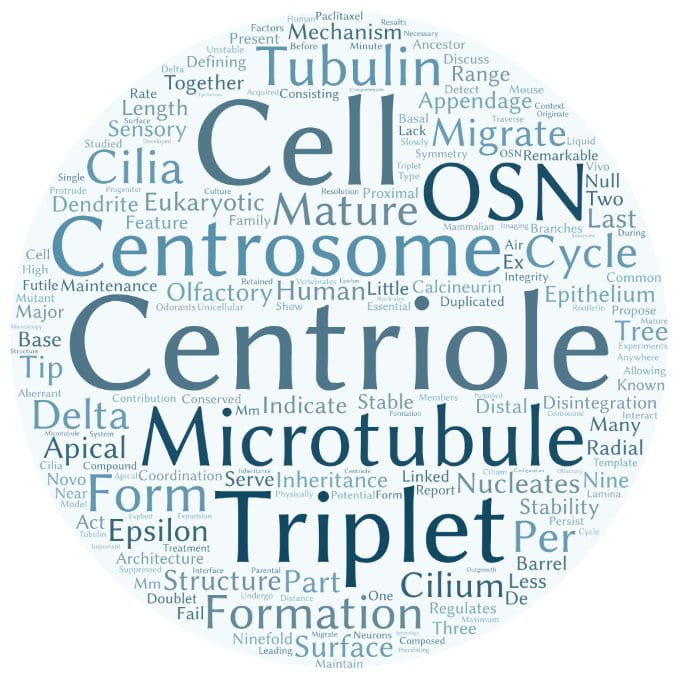
In graduate school, Dr. Wang was fascinated by the way C. elegans worms divided on the microscope, under her very eyes, the same exact way, every single time. She found that she really enjoyed thinking about how cells are organized, and went on to do her postdoc studying the major microtubule organizing center of mammalian cells, the centrosome. The centrosome is found in almost every cell type in human bodies, and defects in its structure or function are related to a wide range of human diseases, including many cancers. Understanding basic cell biological questions about how centrosomes form in human cell lines can lead to understanding some of the things that go wrong in disease.
The lab uses high-resolution microscopy to understand these processes, including a new technique called expansion microscopy, in which you see things beyond the diffraction limit of light by physically expanding the specimen of interest. In other words, they literally make the whole thing bigger. The lab also uses CRISPR/Cas9 gene editing and biochemical techniques.
“Centrosomes and cilia are associated with a bunch of different kinds of diseases, including cancer. In almost every tissue system you can name, there are important roles for centrosomes and their associated organelles called cilia, projections from the cell that either signal or beat or move the cell around. We really want to know how centrosomes are formed, how they act as microtubule organizing centers to organize the rest of the cell, and how they form cilia,” she said.
Learn more about the Wang Lab’s research HERE.